
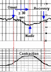
Early deceleration
| A gradual decrease in the fetal heart rate. The time from onset of the deceleration to the lowest heart rate (nadir) during the event is greater than or equal to 30 seconds. The nadir coincides with the peak of the uterine contraction. |
|
Echogenic (hyperechogenic) bowel
Intestine that reflects more sound on an ultrasound examination than usual
making it appear very white. The finding of echogenic bowel is usually a normal
variant in the majority of cases. However, the finding of echogenic bowel has
been associated with an increased risk for chromosomal abnormality, cystic
fibrosis, viral infection (CMV and parvovirus) , unexplained fetal death, growth
restriction, and premature birth.
Echogenic focus
A distinct area that reflects more sound on an ultrasound examination than usual making it appear very white. The term commonly refers to bright spots seen in the ventricles of the heart. Very bright small spots may represent dense papillary muscles or tendons within the heart. Cardiac tumors may also appear as spots within the heart . However, tumors tend to be larger, multiple, and are not as bright as an echogenic focus.
Eclampsia
New-onset convulsions (grand mal seizure) in a woman with preeclampsia. Preeclampsia is a condition characterized by high blood pressure and protein in the urine that develops after the 20th week of pregnancy. The cause of preeclampsia is unknown.
Ectopic Pregnancy
A pregnancy growing outside of the uterus.
Edema
Swelling caused by the accumulation of fluid.
Edwards' syndrome (trisomy 18)
A disorder characterized by severe mental retardation and multiple abnormalities, such as cleft lip and palate, small jaw (micrognathia), low set ears, club feet, clenched fists, intrauterine growth restriction, single umbilical artery, elevated amniotic fluid ( polyhydramnios), and kidney abnormalities. More than 90% of fetuses have a heart defect. The condition is not compatible with life, and only 5% to 10 % of infants survive the first year after delivery.
Edwards' syndrome occurs in about one out of 7500 live births and is caused by extra material from chromosome 18. In most cases (80%) there are three copies of chromosome 18 instead of two. In some cases extra material from a piece of chromosome 18 becomes attached to another chromosome (translocation). Mosaicism (where some of the child's cells have three copies of chromosome 18 and some cells have the normal two copies of chromosome 18) is a less common cause. Recurrence risk is 1% or less for cases caused by three copies of chromosome 18. The risk of having a child with trisomy 18 also increases as a woman gets older. If a parent is a balanced carrier of a translocation, the risk for recurrence is much higher.
Effacement
Thinning or shortening of the cervix.
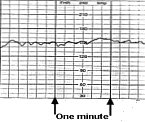
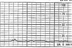
| Electronic fetal monitor
A machine used to make a graphical representation of the fetal heart rate. Most monitors display the fetal heart rate and uterine contractions simultaneous |
 |
|
 |
The ultrasound probe transmits the fetal heart rate in beats per minute. Each small vertical square is 10 beats. Each small horizontal square is 10 seconds. Each large horizontal square is 1 minute. |
 |
. |
The pressure transducer width="149" height="157" transmits the pressure generated by uterine contractions in mm Hg. Each small vertical square is 5 mm Hg. Each small horizontal square is 10 seconds. Each large horizontal square is 1 minute |
 |
Embryo
A fertilized egg from initial cell division until the eighth week of development.
Encephalocele
A defect affecting the skull resulting in the herniation of the meninges and portions of the brain through a bony midline defect in the skull
Engagement
Passage of the widest diameter of the presenting of the part of the fetus (usually the head) to a level below the first sacral vertebra and the symphysis pubis (the joint between the pubic bones at the front of the pelvis).
Epidural
A method of pain relief in which anesthesia is injected into the space around the spinal cord (epidural space) to numb the lower body.
Episiotomy
An incision made between the vagina and rectum to widen the vaginal opening for delivery.
Erythema infectiosum (Fifth disease)
Erythema infectiosum also known as Fifth disease is a common childhood illness caused by a virus called parvovirus B19. The virus appears to be transmitted by person-to-person contact through saliva, sputum, or nasal mucus. About 50% of all adults have been infected sometime during childhood or adolescence.
The incubation period for the infection is 4 to 14 days. Persons infected with the virus may experience fever, malaise, joint pain (particularly of the peripheral joints), and profuse nasal discharge. There may be a lacelike rash on face, trunk , and extremities (See image) .The infection is sometimes mistaken for rubella. Persons are no longer infectious after the appearance of the rash.
Women who become infected with parvovirus for the first time during their pregnancy may pass the virus to their unborn child. Parvovirus can cause severe anemia in the fetus which may lead to congestive heart failure. The heart itself may become enlarged. In addition parvovirus infection has uncommonly been associated with enlarged ventricles in the fetal brain and calcium deposits in the spleen. The overall fetal loss rate related to maternal infection ranges from 2% to 10%.
Women suspected of having a new parvovirus infection during pregnancy are usually followed with serial ultrasounds for 8 to 12 weeks after infection to detect the development of anemia or fetal hydrops.
The thinned out blood of the anemic fetus may be detected by measuring the flow of blood through middle cerebral artery of the brain. Fetal hydrops is a condition where fluid collects in the abdomen, around the lungs, and skin due to congestive heart failure.Leave from the workplace is controversial and is not routinely recommended
Estimated Fetal Weight , EFW
A calculated approximation of the weight of a human conceptus from 70 days' gestational age and older. Where weight is defined as the force that must be applied to an object in order to support it.
EFW can be done by mothers (if they are parous), by clinicians using Leopold manoeuvres or by ultrasound.
EFW may be estimated from Leopolds
mothrer or sonogram get acog and emed citaitons as well.
An ultrasound estimated fetal weight is calculated from models (mathematical equations) using measurements of the fetus obtained during an ultrasound examination, and is sometimes used to predict birth weight. For birth weights in the range of 1000 to 4500 grams models using 3 or 4 fetal biometric indices appear to be more accurate than models that incorporated only 1 or 2 indices. The accuracy of weight estimation decreased at the extremes of BWs, leading to overestimation in low-BW categories as opposed to underestimation when the BW exceeded 4000 g. The precision of most models was lowest in the low-BW groups.
By incorporating the standard ±15% error margin that can be expected with fetal weight estimates made by ultrasonography,
References — Estimated Fetal Weight & Ultrasound Dating
Melamed N, et al. Sonographic fetal weight estimation: which model should be used? J Ultrasound Med. 2009 May;28(5):617-29. PMID: 19389901.
Chauhan SP, Hendrix NW, Magann EF, Morrison JC, Kenney SP, Devoe LD. Limitations of clinical and sonographic estimates of birth weight: experience with 1034 parturients. Obstet Gynecol 1998;91:72–77 (Level II-2).
Chauhan SP, Sullivan CA, Lutton TD, Magann EF, Morrison JC. Parous patients' estimate of birth weight in postterm pregnancy. J Perinatol 1995;15:192–194 (Level II-2).
Ultrasonography is helpful in detecting fetal growth disturbances. Four standard fetal measurements generally are obtained as part of any complete obstetric ultrasound examination after the first trimester: 1) fetal abdominal circumference, 2) head circumference, 3) biparietal diameter, and 4) femur length (41). Fetal morphologic parameters can be converted to fetal weight estimates using published formulas and tables (42). Contemporary ultrasound equipment calculates and displays an estimate of fetal weight on the basis of these formulas. Although all of the published formulas for estimating fetal weight show a good correlation with birth weight, the variability of the estimate generally is plus or minus 16 to 20% (2 standard deviations) (41).
In general, ultrasound-established dates should take preference over menstrual dates when the discrepancy is greater than 7 days in the first trimester and greater than 10 days in the second trimester. Ultrasonography may be considered to confirm menstrual dates if there is a gestational age agreement within 1 week by crown rump measurements obtained in the first trimester or within 10 days by an average of multiple fetal biometric measurements obtained in the second trimester (up to 20 weeks of gestation). Reassigning gestational age in the third trimester should be done with caution because the accuracy of ultrasonography is within 3–4 weeks. Guidelines for assignment of gestational age when a discrepancy exists between menstrual and ultrasound-established dates vary in different ultrasound units.
Ultrasonography in Pregnancy. ACOG Practice Bulletin No. 101. American College of Obstetricians and Gynecologists. Obstet Gynecol 2009;113:451–61.
41. Hadlock FP, Deter RL, Harrist RB, Park SK. Estimating fetal age: computer-assisted analysis of multiple fetal growth parameters. Radiology 1984;152:497–501. (Level II-3).
Estrogen
A group of steroid compounds that includes estradiol, estriol, and estrone. Estrogens are produced by the granulosa cells of developing egg follicles in the ovary, the corpus luteum, and the placenta. They are also produced in small amounts by peripheral tissues, the adrenal glands, and in men by the testes. Estrogens promote the development of secondary female sexual characteristics, and influence the metabolism of bone and the production of many substances produced by the liver.
Estradiol (E2) is the primary hormone produced during a woman's reproductive
years. It reaches high levels just before ovulation occurs during the menstrual
cycle, and is produced in large quantities by the placenta from fetal and
maternal adrenal steroids.
Normal Values
External cephalic version
To manually turn the fetus from a breech (sitting position) presentation to a cephalic presentation (head down nearest to the cervix) by applying external pressure on the mother's abdomen.
Extremely lo birth weight (ELBW)
A birth weight of less than 1000 grams ( 2 pounds 3 ounces)