
Hegar's sign
Softening of the lower uterine segment just above the cervix seen as a probable sign of pregnancy. Originally described by the German gynecologist Ernst Ludwig Alfred Hegar; Hegar's sign may be observed as early as six weeks of pregnancy.
HELLP syndrome
HELLP is an acronym that describes the syndrome of:
H – Hemolysis; EL – elevated liver enzymes; LP – low platelets.
HELLP syndrome usually presents in the third trimester with right upper quadrant or epigastric pain, nausea, and vomiting. HELLP syndrome is considered to be a variant of preeclampsia. However, unlike preeclampsia, hypertension and proteinuria do not need to be present for the diagnosis of HELLP syndrome. HELLP syndrome occurs in approximately 0.2 to 0.6 percent of all pregnancies. The cause of HELLP syndrome is unknown.
Strict criteria for the diagnosis of HELLP syndrome:
- Hemolysis (characteristic peripheral blood smear) and serum lactate dehydrogenase levels > 600 U/L
- Aspartate aminotransferase (AST) and alanine aminotransferase (ALT) elevated more than twice the upper limit of normal.
- Platelet count < 100,000/µL
Complications of HELLP syndrome include abruption, disseminated intravascular coagulation, adult respiratory distress syndrome, pulmonary edema, hepatorenal failure, subcapsular hematoma, and hepatic rupture. Partial HELLP syndrome is also recognized where only one or two features of HELLP syndrome are present.
Hemolytic disease of the newborn (HDN)
Anemia in a newborn infant caused by the destruction of red blood cells. In severe cases jaundice, pallor, an enlarged spleen, or hydrops may be present. The condition is usually caused by an incompatibility between the blood types of the mother and her infant. Antibodies to the following blood groups have been implicated in the development of hemolytic disease of the newborn:
Hemophilia
A group of hereditary disorders characterized by prolonged bleeding and sometimes excessive bleeding. There may be bleeding into joints, gastrointestinal tract, and urinary tract. On laboratory testing, the activated partial thromboplastin time (PTT) is prolonged. However, the prothrombin time (PT) and bleeding time are normal.
Hemophilia A and hemophilia B (Christmas disease) account for most cases of hemophilia. Hemophilia A is caused by a lack of the blood clotting protein factor VIII, and hemophilia B is caused by a lack of the blood clotting protein factor IX. Hemophilia A is approximately 7 times more common than hemophilia B. Both diseases are caused by a defective gene located on the X chromosome.
Since the male contributes the X chromosome to form female and the Y chromosome to form male offspring, all the daughters of a male affected with hemophilia will be carriers of the defective gene. The daughters of a hemophiliac male will pass the gene to 50% of their male offspring, who in turn will have hemophilia.
For the newborn at risk for hemophilia it is best to avoid fetal scalp electrodes, forceps, and vacuum extraction. The pediatrician should be notified of the baby's possible hemophilia. Circumcision, if desired, should be delayed as well as intramuscular injections until coagulation studies are completed and the diagnosis of hemophilia is either established or ruled out.
Hepatitis B (HBV)
A double-stranded DNA virus in the Hepadnaviridae family. Sexual transmission accounts for most adult HBV infections in the United States. Ten to twenty percent of women seropositive for HBsAg transmit the virus to their neonates in the absence of immunoprophylaxis. In women who are seropositive for both HBsAg and HBeAg, vertical transmission is approximately 90%. In patients with acute hepatitis B, vertical transmission occurs in up to 10% of neonates when infection occurs in the first trimester and in 80–90% of neonates when acute infection occurs in the third trimester.
Chronic infection occurs in about 90% of infected infants, 60% of infected children aged < 5 years, and 2%–6% of adults.
The incubation period from time of exposure to onset of symptoms is 6 weeks to 6 months. About one half of acute HBV infections are symptomatic in adults, with 1% of cases resulting in acute liver failure and death. Acutely infected individuals develop loss of appetite, nausea, vomiting, fever, abdominal pain and jaundice.
Among persons with chronic HBV infection, the risk of death from cirrhosis or hepatocellular carcinoma is 15%–25%. HBV infection does not appear to cause birth defects. However, there appears to be a higher incidence of low birth weight and prematurity among infants born to mothers with acute infection during pregnancy.
Hepatitis C (HCV)
Hepatitis C virus (HCV) is a single-stranded RNA virus in the Flaviviridae family. Injecting-drug use currently accounts for 60% of HCV transmission in the United States. Blood transfusion is an uncommon cause of recently acquired infections. Sexual transmission of HCV appears to be inefficient relative to hepatitis B virus (HBV). Transmission between sexual partners of persons with chronic HCV infection with no other risk factors for infection is about 5% (range, 0% to 15%). Household contact with an infected person has been associated with a nonsexual transmission rate of 4% (range, 0% to 11%). Approximately 7–8% of hepatitis C virus–positive women transmit hepatitis C virus to their offspring, with a higher rate of transmission seen in women coinfected with HIV.
The average time to seroconversion after exposure to HCV is 8 to 9 weeks. Acutely infected individuals may develop clinically apparent hepatitis with loss of appetite, nausea, vomiting, fever, abdominal pain and jaundice. Sixty to seventy percent of patients with acute HCV infection are asymptomatic.
Acute HCV infection progresses to chronic HCV infection in most persons (75%–85%). Cirrhosis develops in 10%–20% of persons with chronic hepatitis C and hepatocellular carcinoma in 1%–5%. In one small study acute maternal hepatitis appears to have no effect on the incidence of congenital malformations, stillbirths, abortions, or intrauterine malnutrition. However, acute hepatitis may increase the incidence of prematurity. Pregnancy does not appear to be adversely affected by chronic HCV.
Homocysteine
An amino acid produced during the chemical breakdown of the essential amino acid methionine into cysteine. Homocysteine is also used by the body to regenerate methionine.
Elevated homocysteine levels have been associated with neural tube defects, congenital heart defects, recurrent miscarriage, coronary heart disease, stroke, and peripheral vascular disease.
Hydronephrosis
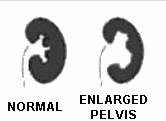
| Enlargement of the renal pelvis (the part of the kidney that collects urine) to greater than 10 mm. Renal pelvis dilation of 4 to 10 mm in anteroposterior diameter is commonly referred to as fetal pyelectasis. The figure at right shows a normal kidney compared to a kidney with minimal hydronephrosis. Dilatation of the urinary tract is detected in utero in 1 per 100 pregnancies. However, only 1 in 500 cases results in significant pathology. |
 |
| Hydronephrosis is usually caused by a blockage of the flow of urine along the urinary tract. Upper urinary tract obstruction is the most common cause of hydronephrosis and may be the result of ureteropelvic junction (UPJ) obstruction, ureteral reflux, or ureterovesical junction obstruction. Less common causes of hydronephrosis include posterior urethral valves, urethral atresia, ectopic ureteroceles, duplication of the collecting system, megacystis–microcolon– intestinal-hypoperistalsis syndrome, and cloacal malformation. | |
Hydrocephaly (hydrocephalus, “water on the brain”)
Enlargement of the spaces within the brain (ventricles) caused by excessive fluid (cerebrospinal fluid). The excessive fluid may cause enlargement of the infant's head.
The abnormally increased fluid may be the result of increased production of fluid, but more commonly is caused by obstruction of fluid flow between the different spaces in the brain. Hydrocephaly has been associated with aqueductal stenosis, spina bifida, X-linked hydrocephalus, Arnold–Chiari malformation, Dandy–Walker malformation, tumors, subarachnoid hemorrhage, infections (CMV and toxoplasmosis), and chromosome abnormalities (8, 9, 13, 15, 18, 21).