Level II / Detailed Ultrasound: What to Expect

A closer look at your baby

Is a Level II ultrasound the same as a detailed ultrasound?

Usually, yes. "Level II ultrasound" is an older name that many offices still use.

Today, doctors often call it a detailed anatomy ultrasound.

It gives the maternal fetal medicine (MFM) team a closer look at your baby.
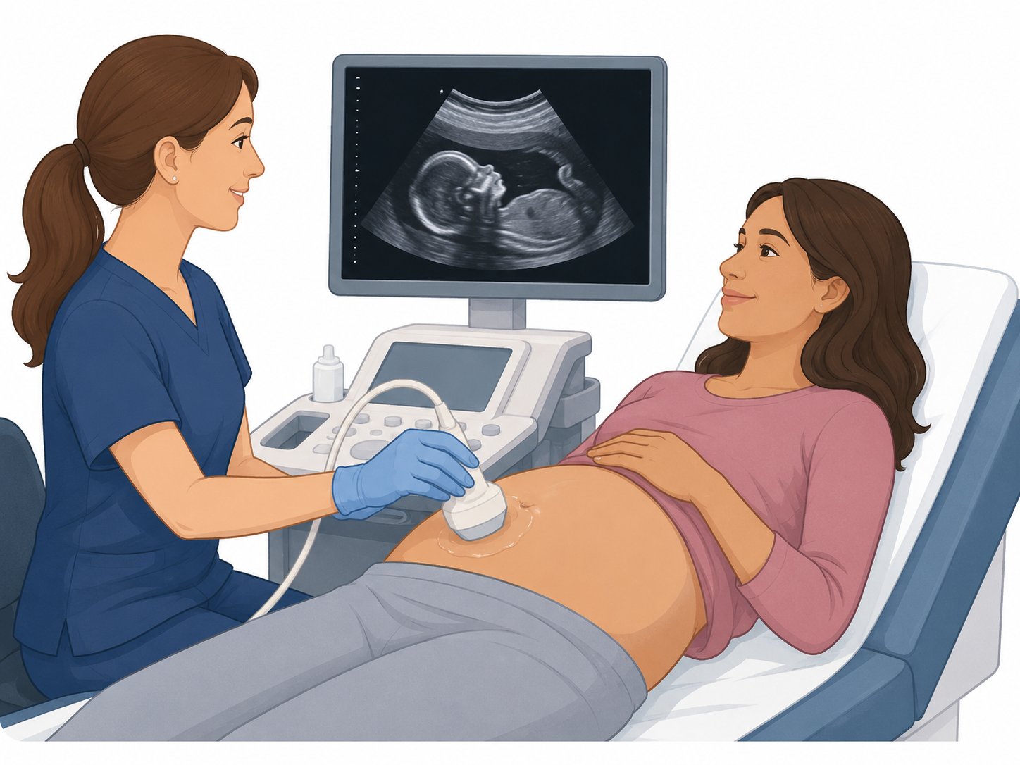
Pregnant patient having an ultrasound while the sonographer moves the probe over the belly.
A detailed ultrasound uses sound waves to make pictures of your baby. It does not use X-rays.
When?Often 18 to 22 weeks
How long?Often 30 to 60 minutes
Radiation?None
Who reviews it?An MFM doctor

How is this different from my OB ultrasound?

Your OB office may check the baby's heartbeat, age, growth, position, and basic body parts.

The MFM scan takes more pictures and measurements when needed.

An MFM doctor reviews the scan.

Who may need a detailed scan?

  • something was seen on another scan
  • a blood test or genetic test raised a question
  • diabetes or another health problem
  • twins or more than one baby
  • a family history or a past pregnancy problem
  • the baby may not be growing as expected

What do we look at?

  • brain and face
  • heart and chest
  • spine
  • stomach, kidneys, and bladder
  • arms, legs, hands, and feet
  • baby's growth
  • placenta, cord, and fluid
  • blood flow or the cervix when needed

What happens during the scan?

  • You lie on an exam table.
  • Gel is put on your belly.
  • A small probe moves over your belly.
  • The ultrasound tech takes many pictures.
  • You may be asked to turn on your side.
  • Sometimes a vaginal ultrasound helps us see the cervix better.

Why might I need more pictures?

Sometimes the baby is hard to see.

The baby may face the wrong way. A hand or foot may block a view.

Coming back for more pictures does not always mean something is wrong.

What if something is found?

The MFM doctor will explain what was found.

You may need another ultrasound scan. You may need a closer look at the baby's heart. You may also meet with a genetic counselor.

Can an ultrasound find every birth defect? No. Some problems are too small to see early in pregnancy. Other problems do not develop or become clear until later. For example, some heart problems, brain changes, kidney problems, bowel blockages, and bone-growth problems may not be seen on an earlier ultrasound. A normal detailed ultrasound is reassuring, but it cannot find every problem. Ultrasound also cannot diagnose a chromosome or gene condition.
Medical references
  1. American Institute of Ultrasound in Medicine (AIUM). Obstetric - Detailed Second/Third Trimester Anatomy Practice Parameter.
  2. American Institute of Ultrasound in Medicine (AIUM). Obstetric - Standard Practice Parameter. Current AIUM standard parameter: 2024.
  3. American College of Obstetricians and Gynecologists (ACOG). Ultrasound Exams. ACOG explains that specialized ultrasound may be used when a problem is suspected because of risk factors or other test results.
  4. International Society of Ultrasound in Obstetrics and Gynecology (ISUOG). Practice Guidelines (updated): Performance of the routine mid-trimester fetal ultrasound scan. Ultrasound Obstet Gynecol. 2022;59:840-856.
  5. International Society of Ultrasound in Obstetrics and Gynecology (ISUOG). Practice Guidelines: performance of third-trimester obstetric ultrasound scan. Ultrasound Obstet Gynecol. 2024;63:131-147.
  6. American Institute of Ultrasound in Medicine (AIUM). Obstetric (Detailed 1st Trimester) Practice Parameter.