Normal thoracic anatomy & standard views

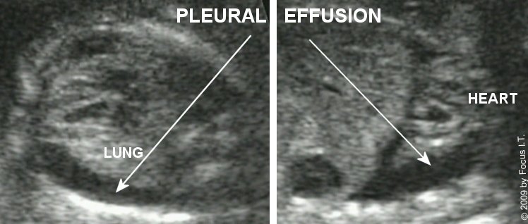
Level II thoracic evaluation focuses on heart position, lung echogenicity, diaphragm continuity, chest shape, and relationship of intrathoracic organs.

Standard thoracic views

  • Four-chamber view (cross-link to cardiac module):
    • Assesses cardiac position (usually left chest with apex pointing left).
    • Evaluates relative lung volumes surrounding the heart.
  • Transverse chest view:
    • Symmetric lung fields of homogeneous, moderately echogenic appearance.
    • Visualization of ribs encircling the chest.
  • Sagittal and coronal views:
    • Continuous, curvilinear diaphragm separating thorax and abdomen.
    • Confirmation of stomach and liver position below the diaphragm.
Normal fetal thorax transverse schematic
Transverse thoracic view: normal lungs, heart position, and diaphragm (schematic placeholder).

Any deviation in organ position (e.g., stomach or liver in the thorax), lung echogenicity, or chest size should prompt detailed evaluation for CDH, CPAM, BPS, effusions, and skeletal dysplasia.

Congenital diaphragmatic hernia (CDH)

Congenital diaphragmatic hernia (CDH) is characterized by a defect in the diaphragm allowing abdominal contents to herniate into the thoracic cavity, compressing the developing lungs and displacing the heart and mediastinum.

Key diagnostic features

Feature Typical findings
Side Most often left-sided; right or bilateral hernias are less common.
Intrathoracic stomach Stomach bubble visualized in the thorax above the diaphragm (classic left-sided CDH sign).
Intrathoracic liver Hypoechoic solid organ in thorax; “liver up” is associated with more severe pulmonary hypoplasia.
Mediastinal shift Heart displaced away from the side of herniation (e.g., to right in left CDH).
Diaphragm Discontinuity or distortion of the normal hemidiaphragm contour.
Table: Summary of typical ultrasound features of congenital diaphragmatic hernia.
Left CDH with stomach and liver up schematic
Left-sided CDH with stomach and part of the liver herniated into the thorax (schematic placeholder).

Lung size and prognostic indices

  • Lung area measurements:
    • Contralateral lung area measured on a standardized transverse view.
    • Used to calculate observed-to-expected lung-to-head ratio (o/e LHR).
  • Observed-to-expected LHR:
    • Observed LHR adjusted for gestational age and expressed as a percentage of expected.
    • Lower values associated with more severe pulmonary hypoplasia and poorer prognosis.
Link to quantitative CDH tools

For detailed o/e LHR and lung area calculations, see:

Associated findings & workup

  • Cardiac position and function (see cardiac module).
  • Other structural anomalies and chromosomal abnormalities.
  • Hydrops or severe mediastinal compression in large hernias.

Prognosis in CDH depends on lung size (e.g., o/e LHR), presence of liver herniation, side of defect, associated anomalies, and access to specialized neonatal and surgical care.

Congenital pulmonary airway malformation (CPAM)

Congenital pulmonary airway malformation (CPAM), formerly “congenital cystic adenomatoid malformation (CCAM),” is a developmental lesion of the lung characterized by abnormal cystic or solid overgrowth of terminal bronchioles.

Key diagnostic features

Feature Typical findings
Location Usually unilateral; often involves a single lobe or segment.
Lesion type Macrocystic (few large cysts), microcystic (many small cysts, solid-appearing), or mixed; echogenicity varies with cyst size.
Mediastinal shift Large lesions can displace the mediastinum and compress contralateral lung.
Vascular supply Typically pulmonary arterial supply and venous drainage; distinguishes from sequestration.
Table: Summary of typical ultrasound features of CPAM.
Macrocystic CPAM schematic
Macrocystic CPAM with multiple large cysts occupying one hemithorax (schematic placeholder).

CPAM Volume Ratio (CVR)

  • Lesion volume often approximated using an ellipsoid formula:
    • Length × width × height × constant (e.g., 0.52).
  • CPAM Volume Ratio (CVR):
    • Lesion volume divided by head circumference.
    • Higher CVR associated with increased risk of hydrops and perinatal morbidity.
Link to CPAM / CVR calculator

For quantitative assessment and serial tracking of lesion size:

Associated findings & evolution

  • Hydrops, polyhydramnios, or mediastinal compression in large lesions.
  • Some CPAMs decrease in size or appear to “disappear” later in gestation, although residual disease can persist postnatally.

Management of CPAM depends on lesion size, CVR, presence of hydrops, and associated anomalies. Options range from expectant management with serial ultrasound to in utero therapy in selected high-risk cases, with postnatal evaluation and resection as indicated.

Bronchopulmonary sequestration (BPS)

Bronchopulmonary sequestration (BPS) is a nonfunctioning mass of lung tissue that lacks normal communication with the tracheobronchial tree and receives systemic arterial supply, usually from the aorta.

Key diagnostic features

Feature Typical findings
Location Often posterior and basal; can be intralobar or extralobar.
Echogenicity Usually homogeneous, solid-appearing echogenic mass.
Arterial supply Systemic arterial vessel arising from aorta or its branches (best seen with color Doppler).
Venous drainage May drain to systemic or pulmonary veins depending on type.
Mediastinal shift Can cause shift and lung compression when large.
Table: Ultrasound features distinguishing BPS from other lung masses.
BPS systemic arterial supply schematic
Bronchopulmonary sequestration with systemic arterial supply from the aorta (schematic placeholder).

Clinical considerations

  • Some lesions remain stable or regress; others contribute to mediastinal shift or hydrops.
  • BPS can coexist with CPAM-like features (hybrid lesions).

Identification of a systemic feeding vessel is key to the diagnosis of BPS. Management is guided by lesion size, hemodynamic impact, presence of hydrops, and associated anomalies, with postnatal surgical planning as needed.

Pleural effusion & hydrothorax

Fetal pleural effusion (hydrothorax) is characterized by fluid accumulation in the pleural spaces, which may be unilateral or bilateral and isolated or part of generalized hydrops.

Sonographic findings

  • Anechoic fluid outlining the lung, often forming a crescent-shaped collection between lung and chest wall.
  • Lungs may appear compressed and centrally displaced.
  • Mediastinal shift and diaphragmatic flattening/eversion in large effusions.
Bilateral fetal pleural effusion schematic
Bilateral pleural effusions compressing the lungs .

Etiologies

  • Primary (isolated) chylothorax.
  • Chromosomal abnormalities and genetic conditions.
  • Infection, cardiac disease, or anemia/hydrops.
  • Associated with other thoracic lesions (e.g., CPAM, CDH) in some cases.

Large pleural effusions may require serial assessment for hydrops, fetal compromise, and consideration of in utero interventions (thoracentesis or shunt) in selected cases, guided by specialized centers.

Lung hypoplasia & small thorax

Lung hypoplasia refers to underdevelopment of the lungs, often secondary to reduced thoracic space or prolonged oligohydramnios.

Common causes

  • Severe, prolonged oligohydramnios (e.g., renal agenesis, severe obstructive uropathy, PPROM).
  • Large thoracic masses (CDH, CPAM, BPS, effusions) compressing the lungs.
  • Severe skeletal dysplasias with small, rigid thorax.

Thoracic size assessment (overview)

  • Chest circumference and thoracic–abdominal circumference ratios.
  • Subjective impression of small, narrow chest with closely spaced ribs.
  • Correlation with limb measurements and overall biometry in skeletal dysplasia.

Prognosis depends on the severity and cause of hypoplasia. Marked reduction in thoracic size and lung volume, especially with underlying skeletal dysplasia or prolonged anhydramnios, is associated with poor postnatal respiratory outcome.

Small chest & chest wall / rib abnormalities

Abnormalities of the chest wall and ribs can result in a small thoracic cage and restrictive lung development, often in the context of skeletal dysplasias.

Sonographic clues

  • Narrow, bell-shaped chest in transverse and coronal views.
  • Shortened ribs or decreased number of visible ribs in a cross-section.
  • Disproportion between chest circumference and abdominal circumference.

Associations

  • Thanatophoric dysplasia and other lethal skeletal dysplasias.
  • Jeune asphyxiating thoracic dystrophy and related conditions.

A small chest in combination with markedly shortened long bones, abnormal bone shape, or other dysmorphic features should prompt evaluation in a skeletal dysplasia context, often with input from genetics and radiology.

Mediastinal and other intrathoracic masses

Intrathoracic masses beyond CPAM and BPS include mediastinal cysts, teratomas, and rarely cardiac or great vessel anomalies presenting as central masses.

Examples

  • Mediastinal teratoma:
    • Complex solid-cystic mass in the anterior mediastinum.
    • May cause mediastinal shift, effusions, and hydrops.
  • Bronchogenic cyst or duplication cyst:
    • Well-circumscribed, anechoic or hypoechoic lesion near the mediastinum.
  • Vascular anomalies:
    • Rarely, anomalous great vessels or aneurysms can simulate mediastinal masses.

When an unusual mediastinal mass is identified, detailed assessment with Doppler, evaluation for effusions and hydrops, and consideration of fetal MRI may help delineate anatomy and guide perinatal planning.

Key thoracic measurements & indices (overview)

Several measurements and ratios aid in assessing fetal thoracic development and the impact of lesions such as CDH, CPAM, and BPS. Detailed numeric calculators are available on dedicated pages; this section summarizes commonly used indices.

Chest circumference

  • Measured on a transverse view of the chest at the level of the four-chamber heart.
  • Compared to gestational age–specific norms and abdominal circumference.

Lung area & LHR (CDH)

  • Contralateral lung area measured in a standardized plane; used to compute LHR and o/e LHR.
  • Lower o/e LHR values indicate more severe pulmonary hypoplasia.

CPAM Volume Ratio (CVR)

  • Ellipsoid lesion volume / head circumference.
  • Higher CVR correlates with increased risk of hydrops and poor outcome.
Link to calculators for thoracic lesions

Thoracic measurements are interpreted in context with overall fetal biometry, presence of hydrops, and other structural anomalies. They aid in counseling, referral to specialized centers, and consideration of fetal interventions.

References

Selected classic and frequently cited references related to fetal thoracic imaging and lung lesions are listed in abbreviated form. Detailed guidelines and lung lesion management protocols should also be consulted.

  1. ISUOG Practice Guidelines for the Sonographic Examination of the Fetal Chest.
  2. Deprest JA, et al. Prenatal diagnosis and management of congenital diaphragmatic hernia.
  3. Jancelewicz T, et al. Fetal lung volume and pulmonary hypoplasia in congenital diaphragmatic hernia.
  4. Adzick NS, et al. Fetal lung lesions: natural history and management of CPAM and BPS.
  5. Crombleholme TM, et al. Congenital cystic adenomatoid malformation volume ratio (CVR) and prediction of outcome.
  6. Wilson RD, et al. Fetal pleural effusion and hydrothorax: diagnosis, natural history, and intervention.
Show expanded citation notes

ISUOG chest guidelines. Outline recommended thoracic planes and documentation for basic and detailed thoracic examinations.

Crombleholme TM, et al. Describe CVR as a prognostic index for CPAM and risk of hydrops.

Deprest JA, et al. Review CDH diagnosis, lung volume assessment, and criteria used for fetal intervention trials.

PMIDs, DOIs, and full citation formatting can be harmonized with other Level II modules according to your preferred reference style on Perinatology.com.