
Diagnosis and Staging of Twin to Twin Transfusion Syndrome
Twin-twin transfusion syndrome is a complication of monochorionic multiple gestation caused by unbalanced net transfusion through placental vascular anastomoses. The usual ultrasound pattern is oligohydramnios in the donor twin and polyhydramnios in the recipient twin. This page summarizes pathophysiology, frequency, definitions, diagnosis, staging, current treatment options, survival counseling, current discussion, surveillance, cervical length and preterm birth risk, and special considerations for monoamniotic twins.
Updated: June 26, 2026. This page is for clinical education and should not replace individualized maternal-fetal medicine consultation, fetal therapy consultation, or local protocols.
On this page
Overview
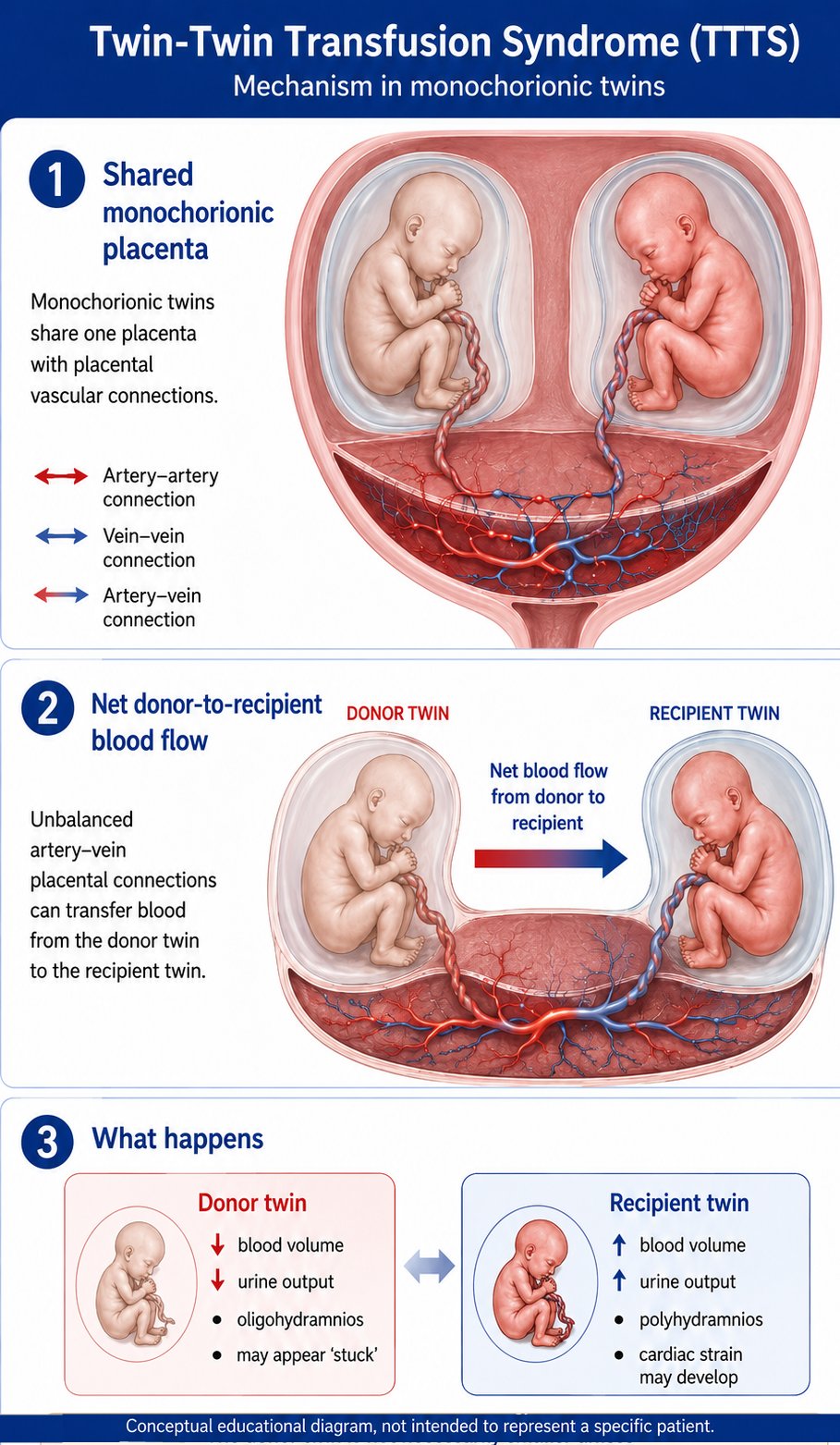
TTTS occurs when intertwin placental vascular connections create a sustained volume shift between fetuses sharing one placenta. The donor twin becomes relatively hypovolemic and oliguric, while the recipient twin becomes relatively hypervolemic, polyuric, and at risk for cardiomyopathy and hydrops.
Mechanism diagram
Pathophysiology and frequency
Placental vascular basis
Most monochorionic placentas contain vascular anastomoses. TTTS develops when net transfusion across deep arteriovenous connections is not adequately balanced by other vascular connections. This produces chronic intertwin volume imbalance rather than a simple difference in fetal size.
Donor physiology
The donor twin loses net circulating volume, develops reduced renal perfusion and urine output, and may show a small or absent bladder, oligohydramnios, fetal growth restriction, abnormal umbilical artery Doppler, or demise.
Recipient physiology
The recipient twin receives excess volume, produces excess urine, and develops polyhydramnios. Cardiovascular strain may lead to ventricular hypertrophy, atrioventricular valve regurgitation, ductus venosus abnormality, hydrops, or demise.
Frequency
TTTS is usually described as affecting approximately 10% to 15% of monochorionic diamniotic twin pregnancies. It is much less common in monochorionic monoamniotic twins, but it can occur.
Definitions
- Monochorionic twins
- Twins sharing one placenta. Chorionicity is the key risk factor for TTTS, TAPS, selective fetal growth restriction, and TRAP sequence.
- Monochorionic diamniotic twins
- Twins sharing one placenta but separated by an intertwin amniotic membrane. This is the usual setting in which classic TTTS fluid criteria are applied.
- Donor twin
- The twin with net transfer of blood volume away from its circulation. The donor is typically oliguric, has a small or absent bladder, and has oligohydramnios.
- Recipient twin
- The twin receiving excess net volume. The recipient is typically polyuric, has a large bladder, has polyhydramnios, and is at risk for cardiac dysfunction and hydrops.
- Stuck twin
- A descriptive ultrasound term for the donor twin compressed against the uterine wall or placenta by severe oligohydramnios. The membrane may be difficult to see, and the fetus may appear to have limited mobility
- Pre-TTTS
- Discordant fluid or evolving findings suspicious for TTTS but not meeting full classic diagnostic criteria. These cases require close short-interval follow-up.
- TAPS
- Twin anemia-polycythemia sequence. This is a monochorionic complication characterized by anemia in one twin and polycythemia in the other, usually diagnosed by discordant MCA-PSV rather than by oligo-polyhydramnios.
Diagnosis
Classic TTTS in monochorionic diamniotic twins is diagnosed when the recipient sac has polyhydramnios and the donor sac has oligohydramnios. A practical reporting approach is to document the deepest vertical pocket or maximum vertical pocket for each sac, donor bladder visibility, Doppler studies, hydrops, growth, and relevant maternal symptoms.
Core ultrasound criteria
- Monochorionic pregnancy.
- Recipient DVP/MVP ≥ 8 cm.
- Donor DVP/MVP ≤ 2 cm.
- Some older or local protocols use a recipient threshold of ≥ 10 cm after 20 weeks; state the threshold used.
Minimum documentation
- Chorionicity and amnionicity.
- Fluid pocket for each twin.
- Donor bladder visible or not visible.
- Umbilical artery, ductus venosus, and umbilical venous Dopplers when feasible.
- Recipient cardiac findings and hydrops.
- Estimated fetal weight and growth discordance.
- MCA-PSV when evaluating for TAPS overlap.
Staging
Quintero staging remains the most common clinical language for TTTS. Staging is based on fluid sequence, donor bladder visibility, Doppler abnormalities, hydrops, and fetal demise.
| Stage | Recipient DVP/MVP ≥ 8 cm and Donor DVP/MVP ≤ 2 cm | Donor bladder | Critical Doppler abnormality | Hydrops | Demise | Clinical meaning |
|---|---|---|---|---|---|---|
| I | Present | Visible | No | No | No | Fluid criteria are met and the donor bladder remains visible. |
| II | Present | Not visible | No | No | No | The donor bladder is not seen during the examination, suggesting marked donor oliguria or anuria. |
| III | Present | Visible or not visible | Yes | No | No | Critically abnormal Doppler is present in either twin. |
| IV | Present | Visible or not visible | May be present | Yes | No | Hydrops is present in one or both twins. |
| V | Present or prior TTTS | Not applicable | Not applicable | Not applicable | Yes | Demise of one or both twins. |
Classic critical Doppler abnormalities include absent or reversed end-diastolic flow in the umbilical artery, reversed ductus venosus a-wave, or pulsatile umbilical venous flow.
2025 Delphi consensus staging proposal - practical reporting additions
The Delphi consensus proposal provides more specific language for evolving disease, donor versus recipient stage III-IV disease, and atypical recipient disease. It is useful for reporting, but it should be identified as a consensus proposal that still needs prospective validation.
| Category | Proposed findings | Practical reporting language |
|---|---|---|
| Pre-TTTS | Discordant fluid not meeting stage I criteria, including isolated polyhydramnios, isolated oligohydramnios, or near-threshold discordance such as MVP ≥ 7 cm with MVP ≤ 3 cm, when not otherwise explained. | Report as pre-TTTS or evolving fluid discordance and recommend follow-up in one week or less. |
| Stage I | Recipient MVP ≥ 8 cm and donor MVP ≤ 2 cm; donor bladder visible; no critical Doppler abnormality; no hydrops. | TTTS stage I. Document symptoms, cervix if relevant, Dopplers, cardiac findings, and trend. |
| Stage II | Same fluid criteria; donor bladder not visible; no critical Doppler abnormality; no hydrops. | TTTS stage II. Urgent fetal therapy referral if in the usual treatment window. |
| Stage III donor | Fluid criteria met and donor has persistent absent/reversed UA end-diastolic flow, absent/reversed DV a-wave, pulsatile UV, or severe tricuspid regurgitation. | TTTS stage III donor. State which Doppler or cardiac abnormality defines the stage. |
| Stage III recipient | Fluid criteria met and recipient has abnormal DV/UV findings, severe tricuspid regurgitation, or other stage III cardiovascular findings. | TTTS stage III recipient. Highlight recipient cardiovascular compromise. |
| Stage IV donor/recipient | Hydrops is present in donor, recipient, or both. | TTTS stage IV donor, stage IV recipient, or stage IV donor plus recipient. |
| Atypical recipient disease | Recipient polyhydramnios and recipient cardiovascular compromise can occur without donor MVP ≤ 2 cm. | Describe as atypical stage III-IV recipient disease or isolated recipient disease and arrange fetal therapy consultation. |
Current treatment options
Treatment depends on gestational age, stage, symptoms, cervical length, technical feasibility, coexisting selective fetal growth restriction or TAPS, and family goals. Fetoscopic laser photocoagulation is the preferred causal therapy for appropriate advanced-stage TTTS because it interrupts the placental anastomoses causing the transfusion imbalance.
| Option | Usual role | Important caveats |
|---|---|---|
| Expectant management | Common for asymptomatic stage I TTTS with at least weekly fetal surveillance. | Consider fetal therapy consultation if symptoms, short cervix, worsening fluid, cardiac findings, or rapid progression. |
| Fetoscopic laser photocoagulation | Standard treatment for stage II-IV TTTS presenting between 16 and 26 weeks when technically feasible. | Requires fetal intervention center. Risks include preterm prelabor rupture of membranes, preterm birth, recurrent TTTS, post-laser TAPS, fetal growth restriction, and fetal loss. |
| Solomon laser technique | Laser approach that coagulates the vascular equator after individual anastomoses are treated, aiming to reduce residual connections. | Technique and outcomes are center dependent; document post-laser surveillance for recurrent TTTS and TAPS. |
| Serial amnioreduction | May be used for symptom relief, later gestational age, or when laser is unavailable or not technically feasible. | Does not correct the vascular cause. It may need repetition and is generally inferior to laser for appropriate severe midtrimester TTTS. |
| Septostomy | Rarely used as primary therapy in contemporary practice. | Can intentionally or unintentionally disrupt the dividing membrane and may complicate subsequent monitoring. |
| Selective reduction / cord occlusion | Individualized option for extreme disease, major anomaly, very poor prognosis for one twin, or when protecting the co-twin is the priority. | Requires detailed counseling at an experienced fetal therapy center. |
| Delivery | Appropriate when gestational age and clinical status make neonatal management safer than continued pregnancy. | For resolved TTTS after laser with dual survivors and no other indication, delivery at 34-36 weeks is generally recommended. |
Survival and prognosis
Outcome counseling should distinguish broad published estimates from the prognosis for the individual pregnancy. Important modifiers include gestational age at diagnosis, Quintero stage, cervical length, placental anatomy, fetal cardiac findings, Dopplers, hydrops, coexisting selective fetal growth restriction, TAPS, preterm membrane rupture, and center experience.
Without effective treatment
Advanced midtrimester TTTS has a high risk of fetal loss and severe neonatal morbidity. Risk is highest with hydrops, fetal demise, severe Doppler abnormalities, very early disease, and marked cervical shortening.
After laser therapy
For stage II-IV TTTS in the usual midtrimester treatment window, fetoscopic laser therapy is the preferred causal treatment when technically feasible. Contemporary counseling often quotes survival of at least one twin in approximately 80% to 90% of treated pregnancies, with dual survival commonly lower and center-specific.
Neurodevelopment
Neurologic risk is influenced by prematurity, fetal demise, hemodynamic instability, stage, and post-procedure complications. Laser therapy reduces morbidity compared with older amnioreduction-only strategies but does not eliminate risk.
| Quintero stage | Survival of at least one twin | No survivor | Counseling note |
|---|---|---|---|
| I | 86.9% | 11.8% | Many cases remain stable or regress, but progression can occur. Management is individualized based on symptoms, cervical length, cardiac findings, and trend. |
| II | 85.0% | 15.0% | Usually managed with urgent fetal therapy evaluation if in the treatment window. |
| III | 81.5% | 18.6% | Doppler or cardiovascular abnormality is present. Prognosis depends on which twin has the abnormality and whether sFGR or recipient cardiac disease coexists. |
| IV | 82.8% | 17.2% | Hydrops is present. Survival of at least one twin can still be possible after laser, but counseling should emphasize fetal and neonatal risk. |
| V | 54.6% | 45.4% | One or both fetuses have demised. Survivor assessment, Dopplers, neurosonography, fetal MRI when appropriate, and individualized delivery planning are needed. |
Discussion: current monochorionic twin pregnancy update
A 2025 Clinical Expert Series review of monochorionic twin pregnancies reinforces that TTTS should be interpreted in the broader context of monochorionic placental disease. TTTS is one of several shared-placenta complications, including TAPS, selective fetal growth restriction, TRAP sequence, monoamnionicity, and single-twin demise. The common theme is that intertwin vascular connections can rapidly change risk for both fetuses.
Screening and referral
The review supports ultrasound surveillance for TTTS every 2 weeks beginning at 16 weeks in monochorionic gestations, with more frequent assessment if isolated polyhydramnios, oligohydramnios, discordant fluid, growth discordance, abnormal Dopplers, or maternal symptoms suggest evolving disease. Referral to a center experienced in fetal therapy is appropriate when fluid or growth discordance is detected or when the diagnosis is uncertain.
Dynamic disease
Most TTTS presents before 26 weeks, with a peak around 20 weeks. Because the disease may evolve quickly, a single reassuring examination does not exclude subsequent progression. Trends in fluid, bladder filling, Dopplers, cardiac findings, growth, and cervical length are often more informative than one isolated measurement.
Stage I dilemma
Stable, asymptomatic stage I TTTS is often managed expectantly with close surveillance, but fetal therapy consultation is reasonable when symptoms, short cervix, cardiac dysfunction, or rapid progression are present. Available randomized data have not shown a clear intact-survival advantage for immediate laser in all stage I cases, but progression can occur and surveillance must be active rather than passive.
Laser and Solomon technique
Fetoscopic laser treats the underlying placental vascular problem by coagulating intertwin anastomoses. The Solomon technique coagulates the placental vascular equator after visible anastomoses are treated, reducing residual connections and lowering recurrent TTTS and post-laser TAPS in published trials.
Periprocedural risk
Important counseling modifiers include gestational age at diagnosis, Quintero stage, progression, preprocedural cervical length, coexisting selective fetal growth restriction, Doppler or cardiac abnormalities, membrane complications, and fetal therapy center experience. Preterm prelabor rupture of membranes remains one of the major risks after fetoscopic laser.
Neurologic and cardiac outcomes
Survival is not the only outcome. Recipient cardiovascular dysfunction, donor placental insufficiency, fetal demise, prematurity, and postprocedure complications all influence neurologic outcome. CNS injury may be present before intervention in some cases, so counseling should include both fetal and neonatal follow-up.
Cervical length and preterm birth risk
Short cervical length is not part of Quintero staging, but it should be documented because it modifies prognosis and fetal therapy counseling. In TTTS, cervical shortening may reflect baseline preterm-birth risk, uterine overdistension from recipient polyhydramnios, contractions, membrane stress, or impending preterm birth.
When to measure
- Measure transvaginal cervical length when TTTS is diagnosed, when symptoms are present, and before fetoscopic laser evaluation when feasible.
- Document cervical dilation, funneling, exposed membranes, contractions, bleeding, or rupture of membranes separately from the cervical length number.
- Cervical length should not be used to change the Quintero stage; it is a preterm-birth and procedural-risk modifier.
What the data suggest
- In laser-treated TTTS, preoperative cervical length below about 25 to 30 mm is associated with higher risk of early preterm delivery.
- One study identified a preoperative cervical length cutoff of 28 mm for spontaneous preterm delivery before 34 weeks after laser surgery.
- In a NAFTNet multicenter cohort of TTTS pregnancies with cervical length <30 mm undergoing fetoscopic laser photocoagulation, 62% delivered before 32 weeks.
Implications
- Short cervix should increase attention to referral timing, perioperative counseling, membrane risk, neonatal counseling, and post-procedure surveillance.
- A very short cervix, progressive shortening, or cervical dilation may affect whether laser, amnioreduction, cerclage, expectant management, or delivery is technically feasible or clinically appropriate.
- Management should be individualized with the fetal therapy center and neonatal team rather than handled as a routine short-cervix pathway.
Intervention caveat
- Evidence is limited and heterogeneous for progesterone, pessary, or cerclage specifically in TTTS with short cervix.
- The NAFTNet cohort did not show a clear improvement in delivery interval, gestational age at delivery, live birth, or neonatal survival with expectant management, vaginal progesterone, pessary, cerclage, or combination therapy.
- SMFM short-cervix guidance recommends against routine progesterone, pessary, or cerclage for cervical shortening in twin gestations outside a clinical trial.
| Finding | Clinical meaning | Suggested action |
|---|---|---|
| Cervical length 30 mm or more | Lower preterm-birth concern relative to short cervix, but TTTS itself remains high risk. | Proceed with TTTS staging and fetal therapy pathway as otherwise indicated. |
| Cervical length 25-29 mm | Borderline or mildly short cervix; risk depends on gestational age, symptoms, and trend. | Document as a prognostic modifier; consider short-interval follow-up and fetal therapy input. |
| Cervical length less than 25 mm | Short cervix; increased concern for preterm birth and procedure-related prematurity. | Include in referral summary and counseling; individualize prevention strategy with fetal therapy/MFM. |
| Cervical length less than 20 mm or dilation | High concern for delivery soon after evaluation, especially with symptoms or exposed membranes. | Urgent fetal therapy/MFM discussion; assess feasibility and timing of intervention, neonatal counseling, and hospital-level resources. |
Surveillance and follow-up
Routine monochorionic diamniotic surveillance
- Determine chorionicity and amnionicity in the first trimester when possible.
- Begin TTTS screening at 16 weeks.
- Repeat ultrasound at least every 2 weeks until delivery when stable.
- At minimum, assess amniotic fluid on both sides of the membrane and fetal bladder filling.
- Ideally include umbilical artery Doppler; consider MCA-PSV from 16 weeks when feasible.
Pre-TTTS or abnormal screen
- Repeat MFM ultrasound in one week or less.
- Increase urgency with maternal symptoms, rapidly rising fluid, short cervix, abnormal Dopplers, hydrops, or early severe growth restriction.
- Follow until findings normalize, stabilize, or progress to TTTS, TAPS, sFGR, or another diagnosis.
After laser therapy
- Weekly surveillance for 6 weeks is commonly recommended.
- If stable, resume every-other-week surveillance unless concerns persist.
- Screen for recurrent TTTS, post-laser TAPS, fetal growth restriction, membrane complications, and fetal demise.
TAPS screening
TTTS staging does not use MCA-PSV, but MCA Doppler is important because TAPS may coexist with TTTS or occur after laser. Prenatal TAPS criteria include donor MCA-PSV > 1.5 MoM with recipient MCA-PSV < 1.0 MoM, or intertwin MCA-PSV difference > 0.5 MoM.
What about monoamniotic twins?
Monochorionic monoamniotic twins share both one placenta and one amniotic cavity. They can develop placental vascular complications, including TTTS, but classic diamniotic TTTS criteria are difficult or impossible to apply because there are not two separate amniotic fluid compartments.
Why diagnosis is different
- No intertwin membrane means there is no donor sac DVP and recipient sac DVP to compare.
- A single common sac may show polyhydramnios, but this does not localize fluid to one fetus.
- The donor may still show a small or absent bladder, growth restriction, abnormal Doppler, or anemia.
- The recipient may show a large bladder, cardiomegaly, valve regurgitation, ductus venosus abnormality, hydrops, or polycythemia.
Implications
- Quintero staging should not be used without a clear caveat in monoamniotic twins.
- Refer suspected cases to a fetal therapy center because diagnosis and treatment are individualized.
- Assess for competing monoamniotic complications, including cord entanglement, cord compression, TRAP sequence, structural anomaly, selective fetal growth restriction, and TAPS.
- Laser therapy may be considered in selected cases but can be technically more complex because both fetuses and both cords are in one sac.
Differential diagnosis and common mimics
Selective fetal growth restriction
sFGR may coexist with TTTS. Donor stage III disease may overlap with abnormal umbilical artery Dopplers from placental insufficiency.
TAPS
TAPS produces anemia-polycythemia without classic oligo-polyhydramnios. MCA-PSV discordance is central to screening and diagnosis.
Atypical recipient disease
Recipient cardiovascular compromise may occur without donor oligohydramnios meeting classic stage I criteria. Describe the recipient cardiac and Doppler findings clearly.
Discordant anomaly
Renal disease, bladder outlet obstruction, cardiac disease, genetic syndromes, or structural anomalies can cause discordant fluid or growth.
Membrane rupture or maternal disease
PPROM, infection, placental insufficiency, diabetes, or other maternal disease can alter fluid and growth patterns and should be considered when findings are atypical.
TRAP sequence
TRAP is a monochorionic complication with a pump twin supporting an acardiac twin. It is not TTTS but may coexist in the differential of monochorionic hemodynamic disease.
References and linked sources
References are placed in a collapsible section for easier phone viewing.
- Miller RS, Miller J, Obican S, Simpson L; Society for Maternal-Fetal Medicine Publications Committee. Society for Maternal-Fetal Medicine Consult Series #72: Twin-twin transfusion syndrome and twin anemia-polycythemia sequence. Am J Obstet Gynecol. 2024. PubMed | SMFM
- Khalil A, Sotiriadis A, Baschat A, et al. ISUOG Practice Guidelines (updated): role of ultrasound in twin pregnancy. Ultrasound Obstet Gynecol. 2025;65:253-276. PubMed | Journal
- Simpson LL. Update on Management and Outcomes of Monochorionic Twin Pregnancies. Obstet Gynecol. 2025;145:486-502. doi:10.1097/AOG.0000000000005891. DOI
- Di Mascio D, Khalil A, D'Amico A, Buca D, Benedetti Panici P, Flacco ME, Manzoli L, Liberati M, Nappi L, Berghella V, D'Antonio F. Outcome of twin-twin transfusion syndrome according to Quintero stage of disease: systematic review and meta-analysis. Ultrasound Obstet Gynecol. 2020;56:811-820. PubMed | DOI
- Papanna R, Mann LK, Baschat AA, Bebbington MW, Khalek N, Johnson A, et al. Cervical length in prediction of preterm birth after laser surgery for twin-twin transfusion syndrome. Ultrasound Obstet Gynecol. 2015;45:175-182. PubMed | DOI
- Buskmiller C, Bergh EP, Brock C, Miller J, Baschat A, Galan H, et al. Interventions to prevent preterm delivery in women with short cervix before fetoscopic laser surgery for twin-twin transfusion syndrome. Ultrasound Obstet Gynecol. 2022;59:169-176. PubMed | DOI
- Biggio J; Society for Maternal-Fetal Medicine Publications Committee. SMFM Consult Series #70: Management of short cervix in individuals without a history of spontaneous preterm birth. Am J Obstet Gynecol. 2024;231:B2-B13. SMFM | PubMed
- Bartin R, Andrioli Peralta CF, Peneluppi Horak AC, Rodrigues da Costa KJ, Colmant C, Stirnemann J, et al. Management of short cervix in twin-to-twin transfusion syndrome: a role for pessary placement following fetoscopic laser surgery? Am J Obstet Gynecol. 2024;230:91.e1-91.e12. PubMed | DOI
- Paek B, Walker M, Lewi L, Slaghekke F, Obican S, Papanna R, et al. Consensus update on twin-twin transfusion syndrome staging: An international Delphi study. Pregnancy. 2025;1:e70107. DOI
- Quintero RA, Morales WJ, Allen MH, Bornick PW, Johnson PK, Kruger M. Staging of twin-twin transfusion syndrome. J Perinatol. 1999;19:550-555. PubMed
- Senat MV, Deprest J, Boulvain M, Paupe A, Winer N, Ville Y. Endoscopic laser surgery versus serial amnioreduction for severe twin-to-twin transfusion syndrome. N Engl J Med. 2004;351:136-144. PubMed
- Slaghekke F, Lopriore E, Lewi L, et al. Fetoscopic laser coagulation of the vascular equator versus selective coagulation for twin-twin transfusion syndrome: an open-label randomized controlled trial. Lancet. 2014. PubMed
- van der Veeken L, Stirnemann J, Simpson L, et al. Laser for twin-to-twin transfusion syndrome: a guide for endoscopic surgeons. Prenat Diagn. 2020. PMC
- Murgano D, Prefumo F, Fichera A, et al. Outcome of twin-to-twin transfusion syndrome in monochorionic monoamniotic twin pregnancies. Ultrasound Obstet Gynecol. 2020. PubMed
- Khalil A, Gordijn S, Ganzevoort W, et al. Consensus diagnostic criteria and monitoring of twin anemia-polycythemia sequence. Ultrasound Obstet Gynecol. 2020. Journal
- National Institute for Health and Care Excellence. Twin and triplet pregnancy: NICE guideline NG137. NICE recommendations
- ISUOG VISUOG. Twin-Twin Transfusion Syndrome. ISUOG VISUOG